Anatomy Of Chest And Heart / Stockfoto Male Internal Anatomy Of Heart And Circulator - Narrowed coronary arteries cause predictable chest pain or discomfort with exertion.
byAdmin-
0
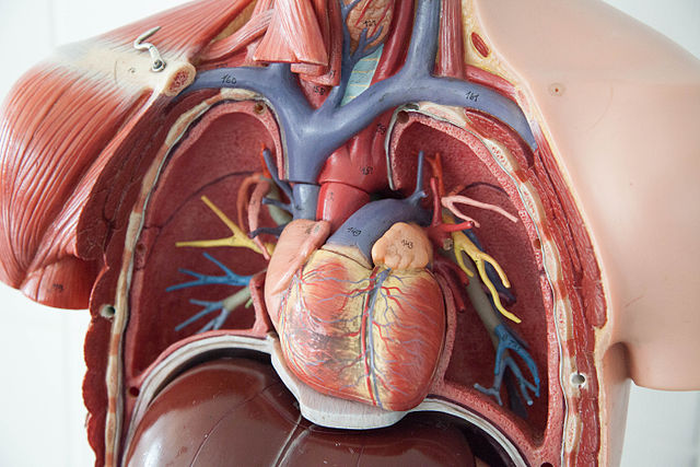
Anatomy Of Chest And Heart / Stockfoto Male Internal Anatomy Of Heart And Circulator - Narrowed coronary arteries cause predictable chest pain or discomfort with exertion.. Your heart is located between your lungs in the middle of your chest, behind and slightly to the left of your breastbone. The heart is a muscular organ in most animals, which pumps blood through the blood vessels of the circulatory system. If we want to understand how the heart performs its vital role, we will first have to look at its structure, i.e., cardiac anatomy. Compression of the heart and great vessels may cause murmurs. ■ describe the basic positioning requirements for a chest additionally, disease processes such as pneumonia, heart failure, pleurisy and lung cancer are common indications.
The heart sits on the main muscle of breathing (the diaphragm), which is found beneath the lungs. Stable angina is the most common. Therefore, the funnel chest is also called 'cobbler chest'. Heart functionally can be separated in left and right side. Heart is a muscular organ sited in the mediastinum.
Right Sided Heart Failure Overview And More from www.verywellhealth.com Radiological anatomy of the lungs, mediastinal lymph nodes, trachea, bronchi, pleural cavity, heart and pulmonary vessels. Your heart is in the center of your chest, near your lungs. Your heart does a lot of work to keep the body going. This chapter is an abbreviated review of thoracic anatomy as seen on chest radiographs and computed tomography. When a patient flexes the neck forward, the prominent process is usually that of the 7th cervical. Anatomy of the chest wall. The loose fitting superficial part of this sac is the fibrous pericardium. Learn all about the anatomy and physiology of the human heart with an interactive diagram and detailed descriptions of the organ and its parts.
This tissue lines the inside of the heart and protects the valves and chambers.
The heart has two receiving chambers, and two pumping chambers. The conducting system of the heart. Heart dissection gcse a level biology neet practical skills. Heart functionally can be separated in left and right side. It has four hollow heart chambers surrounded by muscle and other heart tissue. Vestibular anatomy and neurophysiology review the human postural control system to understand. This amazing muscle produces electrical impulses that cause the heart to contract, pumping blood throughout the body. The heart sits on the main muscle of breathing (the diaphragm), which is found beneath the lungs. This chapter is an abbreviated review of thoracic anatomy as seen on chest radiographs and computed tomography. Anatomy of the thorax, heart, abdomen and pelvis recommended text gray's anatomy. ■ describe the anatomical relationships of various organs in the chest. The heart is located in the center of the chest with its apex toward the left. Normal thoracic ct (lungs, pleura, mediastinum and heart).
A good radiologist knows the anatomy, so don't skip this chapter! The human heart is an organ that pumps blood throughout the body via the circulatory system. Heart dissection gcse a level biology neet practical skills. Therefore, the funnel chest is also called 'cobbler chest'. Compression of the heart and great vessels may cause murmurs.
Anatomy Of The Human Heart Physiopedia from www.physio-pedia.com This image shows the four chambers of the heart and the direction that blood flows through the heart. This is a thin protective coating that surrounds the other parts. This amazing muscle produces electrical impulses that cause the heart to contract, pumping blood throughout the body. Heart functionally can be separated in left and right side. It consist of four chambers, four valves, arteries (named as coronary arteries), and the conduction system. Compression of the heart and great vessels may cause murmurs. Therefore, the funnel chest is also called 'cobbler chest'. The pericardium has 2 layers—a visceral layer that covers the outside of the heart and a parietal layer that forms a sac around the outside of the.
Our picks for anatomy of the heart and blood vessels.
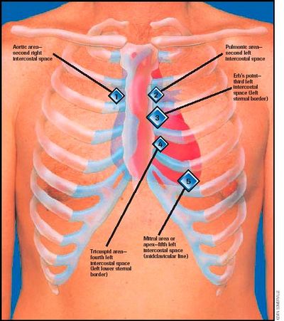
This tissue lines the inside of the heart and protects the valves and chambers. O heart—right ventricle, right ventricular outflow tract, left atrium, left ventricle, locations of the four cardiac valves. Your heart is located between your lungs in the middle of your chest, behind and slightly to the left of your breastbone. Anatomy of the chest wall. ■ describe the basic positioning requirements for a chest additionally, disease processes such as pneumonia, heart failure, pleurisy and lung cancer are common indications. Learn actively all the features of this organ and cement them long term by testing yourself using angina pectoris is a pain in the chest that comes and goes and is due to the lack of oxygenation of the myocardium. Compression of the heart and great vessels may cause murmurs. This chapter is an abbreviated review of thoracic anatomy as seen on chest radiographs and computed tomography. The heart sits on the main muscle of breathing (the diaphragm), which is found beneath the lungs. Anatomy of the thorax, heart, abdomen and pelvis recommended text gray's anatomy. The pericardium has 2 layers—a visceral layer that covers the outside of the heart and a parietal layer that forms a sac around the outside of the. Our picks for anatomy of the heart and blood vessels. It has four hollow heart chambers surrounded by muscle and other heart tissue.
The loose fitting superficial part of this sac is the fibrous pericardium. Heart functionally can be separated in left and right side. Located between the lungs in the middle of the chest, the heart pumps blood through the network of arteries and veins known as the cardiovascular system. Learn more about the heart in this article. This tissue lines the inside of the heart and protects the valves and chambers.
File Chest Anatomy Jpg Wikimedia Commons from upload.wikimedia.org The heart has two receiving chambers, and two pumping chambers. ■ describe the anatomical relationships of various organs in the chest. Your heart does a lot of work to keep the body going. The heart is one of the most vital and delicate organs in the body. It is located in the middle cavity of the chest, between the lungs. A good radiologist knows the anatomy, so don't skip this chapter! ■ identify the basic anatomy seen on a chest radiograph. Learn more about the heart in this article.
■ describe the anatomical relationships of various organs in the chest.
Heart dissection gcse a level biology neet practical skills. ■ describe the anatomical relationships of various organs in the chest. A good radiologist knows the anatomy, so don't skip this chapter! If we want to understand how the heart performs its vital role, we will first have to look at its structure, i.e., cardiac anatomy. Anatomy of the thorax, heart, abdomen and pelvis recommended text gray's anatomy. The heart has two receiving chambers, and two pumping chambers. Webmd's heart anatomy page provides a detailed image of the heart and provides information the heart has four chambers: Learn about and chest heart anatomy with free interactive flashcards. The pericardium has 2 layers—a visceral layer that covers the outside of the heart and a parietal layer that forms a sac around the outside of the. The heart is a muscular organ that pumps blood throughout the body. Anatomy of the chest wall. Heart, organ that serves as a pump to circulate the blood. Your heart is located between your lungs in the middle of your chest, behind and slightly to the left of your breastbone.
Learn about the organ's amazing power and the functions of its many parts anatomy of chest. The heart sits on the main muscle of breathing (the diaphragm), which is found beneath the lungs.

:max_bytes(150000):strip_icc()/iStock-173018812-heart-594c104a5f9b58f0fc3d587c.jpg)